Superstructure-dependent Decoherence of Surface Electrons on Ge (001)
K. Nakatsuji and F. Komori
Electron loses its phase memory when it is inelastically scattered. The way of decoherence depends on the scattering mechanism. On metal and semiconductor surfaces, the observed decay of electronic standing waves is attributed to the decoherence, and is determined by electron-phonon or electron-electron scattering. We have studied the decoherence of the surface state electrons at the clean Ge(001) surface, where two stable superstructures with almost the same quasi one-dimensional (1D) electronic state coexist at 80 K.
The surface consists of buckled Ge dimers, which align in the [110] direction and form dimer rows. The empty π*-band made of the dangling bond states of the surface Ge dimers behaves like a 1D free electron along the dimer row. There are two stable superstructures, p(2x2) and c(4x2) (see Fig. 1), and we can control the surface structure reversibly between them with hysteresis by changing the sample bias voltage VB for the surface observation using scanning tunneling microscopy (STM). [1] We measured the decay of the π*-band standing waves from monoatomic steps in differential conductance dI/dV images after preparing a surface with the both superstructures by the VB change.
Figure 1(a,b) shows the topographic and dI/dV images of the same area. On this surface, both p(2x2) and c(4x2) structures coexist on the lower terrace of a single step [1]. We can see that the sanding waves from the step decay in several nm in Fig. 1 (b). The cross sections of the differential conductance image from the step are plotted in Figs. 2 for VB = 0.4 and 0.5 V on the p(2x2) and c(4x2) areas. We fitted each standing wave to the equation: dI/dV(x) = Aexp(-2x/Lc)sin(2kx +δ). Here the fitting parameter Lc is the decay length of the wave, A and δ the amplitude and phase shift of the wave, and k the wave number of the π* electron. The dispersion relation of the π*-band for each surface obtained by this analysis is almost common to the both surfaces. Whereas, Lc on the p(2x2) surface is longer than on the c(4x2) for both bias voltages. This suggests that the superstructure-dependent electron-phonon interaction in the two surfaces makes the decoherence of the surface electrons different.
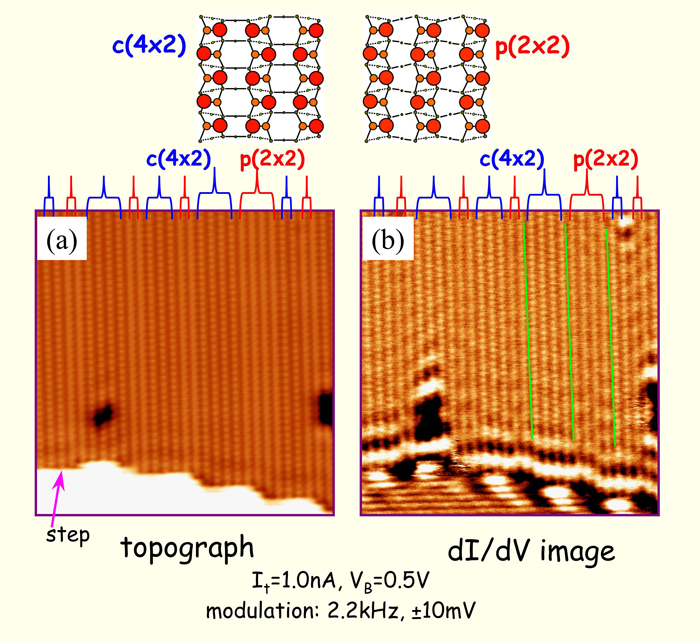
Fig. 1 Topographic (a) and dI/dV (b) images of a clean Ge(001) surface at 80 K. Two superstructures, c(4x2) and p(2x2) shown schematically in the upper panels, were intentionally positioned at the lower terrace of the step by a surface manipulation [1]. Standing waves of the π* electrons from the step edge are seen in (b).
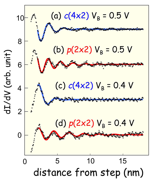
Fig. 2 Cross sections of the standing waves in the dI/dV images for c(4x2) (a,c) and p(2x2) (b,d) areas. [1] The sample bias voltage VB is 0.5 V (a,b) or 0.4 V (c,d). The wave length increases with increasing VB. Blue and red curves are damping oscillation curves fitted to the data. The decay length of the wave for the same bias voltage is shorter on the c(4x2) surface than on the p(2x2) surface.
References
1. Y. Takagi, Y. Yoshimoto, K. Nakatsuji and F. Komori, J. Phys. Soc. Jpn. 72, 2425 (2003); Surf. Sci. 559 1(2004); Phys. Rev. B75 115304 (2007).